Revolutionary 3D-Printed Brain Sensors: A New Era in Neural Interfaces
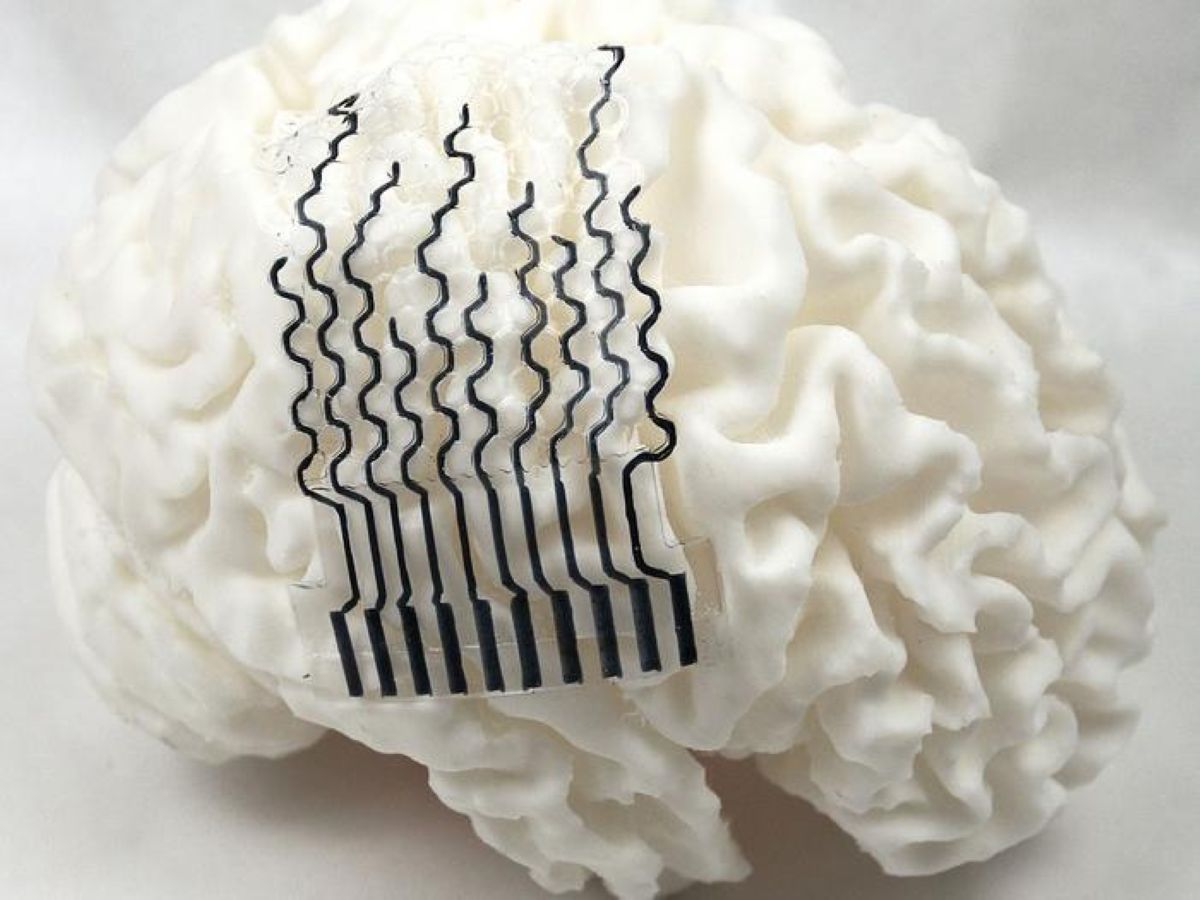
A groundbreaking innovation in the field of neuroscience has led to the development of 3D-printed brain sensors that can be tailored to an individual's unique neural map. This breakthrough has the potential to revolutionize the way we approach neural interfaces, particularly in the treatment and monitoring of neurodegenerative diseases. Researchers at Penn State have successfully created soft, stretchable bioelectrodes that can conform to the intricate ridges and grooves of the brain, providing unparalleled connectivity and signal quality.
The human brain is a complex and highly individualized organ, with no two brains sharing the exact same structure. Despite this, traditional neural implants have typically relied on a one-size-fits-all design, which can lead to suboptimal performance and potentially damaging side effects. The new 3D-printed sensors, designed to match the specific geometry of a patient's brain, offer a significant improvement over these outdated methods. By using a honeycomb-inspired design, the researchers have created electrodes that are not only flexible and strong but also biologically compatible and gentle on the brain tissue.
The development of these innovative sensors is based on a deep understanding of the brain's complex structure and function. The cerebral cortex, the outer layer of the brain, is characterized by a series of folds and grooves that allow for efficient communication between different regions. The process of gyrification, which occurs during fetal development, shapes the brain into its unique configuration, with the resulting gyri and sulci varying significantly from person to person. By taking into account these individual differences, the researchers have created electrodes that can be tailored to the specific needs of each patient.
The production process begins with an MRI scan of the patient's brain, which is then used to create a detailed simulation of the brain's structure. This information is used to design and 3D-print the electrodes, which are made from a water-rich material known as hydrogel. The honeycomb structure of the electrodes provides a unique combination of flexibility, strength, and cost-effectiveness, making them an attractive solution for a wide range of neural interface applications.
The implications of this breakthrough are far-reaching and exciting. Neural interfaces have the potential to revolutionize the treatment of neurodegenerative diseases such as Parkinson's, Alzheimer's, and epilepsy, as well as provide new opportunities for prosthetic control and brain-computer interfaces. By providing a more precise and gentle means of interacting with the brain, these innovative sensors may help to unlock new avenues of research and treatment, leading to improved outcomes and enhanced quality of life for millions of people around the world.
The researchers' use of finite element analysis, a computational method that simulates the behavior of complex systems, has enabled them to optimize the design of the electrodes and predict their performance in different scenarios. This multidisciplinary approach, combining materials science, mechanical engineering, and neuroscience, has yielded a truly innovative solution that has the potential to transform the field of neural interfaces.
Summary Points
3D-printed brain sensors can be tailored to an individual's unique neural map, providing unparalleled connectivity and signal quality
The honeycomb-inspired design of the electrodes offers a unique combination of flexibility, strength, and cost-effectiveness
The production process begins with an MRI scan of the patient's brain, which is used to create a detailed simulation of the brain's structure
The electrodes are made from a water-rich material known as hydrogel, which is biologically compatible and gentle on the brain tissue
The implications of this breakthrough are far-reaching and exciting, with potential applications in the treatment of neurodegenerative diseases and the development of brain-computer interfaces